Our Services

Discover our cutting-edge medical imaging services, including Digital X-ray, IVP for urinary system imaging, HSG for female reproductive health, Barium Studies for the GI tract, DOPG for oral health, Digital Mammography, Doppler for Pregnancy, Ultrasound, Fibroscan for Liver, Echocardiography, ECG, MRI, Angiography, MRCP for bile ducts and pancreas, Venography, and CT scans. We provide accurate and comprehensive diagnostics for all your healthcare needs.

X-ray services

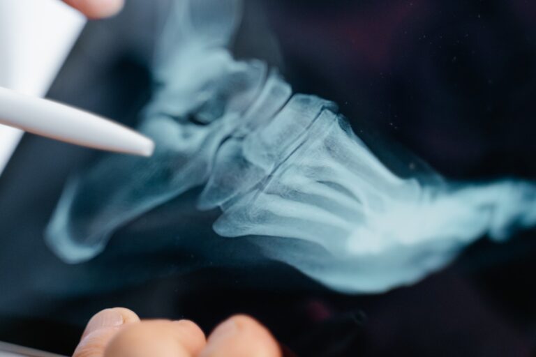
Digital X-ray,

Also known as digital radiography, is a modern imaging technique that uses digital technology to capture and process X-ray images. Traditional X-ray imaging involves exposing a film to X-ray radiation, which produces an image based on the varying absorption of X-rays by different tissues in the body. In contrast, a digital X-ray uses a digital detector to capture the X-ray information, which is then converted into an electronic signal and displayed on a computer screen.
IVP

An Intravenous Pyelogram (IVP), also known as an intravenous urogram (IVU), is a medical imaging procedure that uses digital X-ray technology to visualize the urinary system, specifically the kidneys, ureters, and bladder. It involves the use of a contrast dye that is injected into a vein, typically in the arm. The contrast dye helps highlight the urinary tract structures, allowing for the identification of various conditions and abnormalities.

HSG

A Hysterosalpingography (HSG) is a medical imaging procedure that involves using X-rays to examine the uterus and fallopian tubes. It's often used to diagnose issues related to female reproductive health, such as infertility, abnormal uterine bleeding, and suspected tubal blockages. The procedure helps visualize the shape and structure of the uterine cavity and the patency (openness) of the fallopian tubes.

Barium Study

A barium study, also known as a barium contrast examination or barium swallow, is a medical imaging procedure used to visualize the upper gastrointestinal (GI) tract. It involves the use of barium sulfate, a radiocontrast agent, which is a white, chalky substance that is visible on X-ray images. The procedure helps to diagnose various conditions and abnormalities in the esophagus, stomach, and small intestine.

Retrograde Urethrography (RGU)

RGU is commonly used when there is suspected urethral trauma, narrowing (stricture), or other urethral issues.RGU is primarily used to examine the male urethra, although it can also be performed in females in specific cases. During an RGU, a contrast agent (dye) is injected into the urethra through a thin catheter. X-ray images are then taken as the contrast flows through the urethra, outlining its structure. This helps in identifying any abnormalities, such as strictures, obstructions, or injuries.

RGU" and "MCU" are abbreviations for radiological imaging tests that focus on the urinary tract. They stand for Retrograde Urethrography (RGU) and Micturating Cystourethrogram (MCU). These tests are used to evaluate the structure and function of the urinary system, particularly the urethra, bladder, and sometimes the ureters. Here's what each of these tests involves:

Micturating Cystourethrogram (MCU)

MCU is often performed in children to evaluate vesicoureteral reflux (VUR), a condition where urine flows backward from the bladder into the ureters. It can also be used to investigate recurrent urinary tract infections. MCU is used to assess the bladder and the lower urinary tract, including the urethra.During an MCU, a catheter is inserted into the bladder, and a contrast agent is instilled into the bladder. The patient is then asked to urinate while X-ray images are taken in real time. This allows the radiologist to observe the flow of contrast through the urethra and into the bladder, assessing bladder function and detecting any reflux (backward flow of urine) from the bladder into the ureters.

Ntnb Scan

The “Nb NT scan” is a medical procedure known as the “Nuchal Translucency (NT) scan,” The NT scan is typically performed between the 11th and 14th weeks of pregnancy, which corresponds to the first trimester to assess the risk of certain chromosomal abnormalities in the developing fetus, particularly Down syndrome (Trisomy 21) and other genetic conditions. The primary purpose of the NT scan is to measure the thickness of the nuchal translucency, a small fluid-filled space at the back of the fetus’s neck, using ultrasound imaging.An increased nuchal translucency thickness can be an early indicator of certain chromosomal abnormalities.

Digital OPG

A Digital Orthopantomogram (DOPG), also known as a panoramic dental X-ray or OPG is a specialized dental imaging technique used to capture a wide and detailed view of the entire oral and maxillofacial region, including the teeth, jaws, and surrounding structures. It’s a valuable tool for diagnosing dental and maxillofacial issues, planning treatments, and evaluating the overall oral health of a patient.

Digital Mammography

Digital mammography is a specialized imaging technique used to screen for and diagnose breast cancer and other breast-related abnormalities. It’s an advanced version of traditional mammography that uses digital technology to capture and store X-ray images of the breast tissue. Digital mammography offers several advantages over film-based mammography, including improved image quality, faster image acquisition, and the ability to manipulate and enhance images for more accurate diagnosis.

Ultrasound

Ultrasound, also known as sonography, is a medical imaging technique that uses high-frequency sound waves to create real-time images of the inside of the body. It is a non-invasive and safe diagnostic tool that is widely used in various medical specialties, including obstetrics, cardiology, radiology, and more.
3D Ultrasound

A 3D ultrasound is a medical imaging technique that uses ultrasound technology to create three-dimensional images of a developing fetus in the womb. They do not use ionizing radiation, such as X-rays. Like traditional 2D ultrasound, a 3D ultrasound uses high-frequency sound waves to create detailed and realistic images of the fetus during pregnancy. It allows for a clearer view of the baby's features and can be used for both medical and bonding purposes. They can show facial features, fingers, toes, and other details more clearly.They can show facial features, fingers, toes, and other details more clearly.

TVS

TVS stands for "Transvaginal Ultrasound." It is a medical imaging procedure that involves, examines and assesses various gynecological conditions and concerns. It uses ultrasound technology to create images of the female reproductive organs, particularly the uterus and ovaries. It provides detailed images of the pelvic region, aiding in the diagnosis and monitoring of reproductive health issues.TVS can be performed at various stages of the menstrual cycle and is often used when a healthcare provider suspects or needs to investigate a gynecological issue. The results of a TVS can help guide further treatment or interventions as needed, depending on the findings.

Level-2

A Level-2 ultrasound, also known as a "second-trimester ultrasound" or a "detailed anatomy scan," is a type of prenatal ultrasound performed during the second trimester of pregnancy, typically between 18 to 22 weeks.The primary purpose of a Level-2 ultrasound is to assess the development and health of the fetus in more detail. It provides a comprehensive examination of the fetal anatomy and can help detect structural abnormalities, birth defects, or other health concerns.During a Level-2 ultrasound, various aspects of the fetus are evaluated, including the head, brain, heart, spine, limbs, organs, and other structures. The examination provides detailed images and measurements to ensure the baby's well-being and to identify any potential issues that may require medical attention.

Doppler

Doppler ultrasound is a specialized ultrasound technique that measures blood flow and evaluates the movement of blood within blood vessels. Doppler ultrasound is used in various medical specialties, including obstetrics, cardiology, and vascular medicine. It can help assess blood circulation, detect abnormalities in blood vessels, and monitor conditions such as deep vein thrombosis, carotid artery disease, and fetal well-being during pregnancy. The Doppler ultrasound technique is based on the Doppler effect, which involves changes in the frequency of sound waves reflected off moving objects (in this case, blood cells). By analyzing these frequency changes, healthcare providers can determine the direction and speed of blood flow.

Fibroscan for Liver

FibroScan is a non-invasive medical procedure used to assess the degree of fibrosis (scarring) in the liver. It’s a specialized technology that provides a quantitative measurement of liver stiffness, which is a key indicator of fibrosis severity. The procedure is commonly used to evaluate the health of the liver in individuals

Echocardiography

Echocardiography, often referred to as an “echo,” is a medical imaging technique that uses sound waves (ultrasound) to create real-time images of the heart. It’s a non-invasive and painless procedure that provides valuable information about the structure and function of the heart. Echocardiography is commonly used to diagnose and monitor various heart conditions and is an essential tool in cardiology.

ECG

An electrocardiogram (ECG or EKG) is a medical test that records the electrical activity of the heart over a period of time. It is a commonly used diagnostic tool to assess the heart’s rhythm, rate, and overall electrical health. ECGs are used to detect various heart conditions, including arrhythmias (irregular heart rhythms), heart attacks, and other cardiac abnormalities.

MRI

Magnetic Resonance Imaging (MRI) is a powerful medical imaging technique that uses a strong magnetic field, radio waves, and computer technology to create detailed and cross-sectional images of the body’s internal structures. MRI provides valuable information about organs, tissues, bones, and various medical conditions.
Angiography

Angiography, also known as arteriography, is a medical imaging technique used to visualize blood vessels and blood flow within the body. It involves the use of contrast dye and X-ray technology to create detailed images of blood vessels, including arteries, veins, and capillaries. Angiography is commonly used to diagnose and assess various vascular conditions, such as blockages, aneurysms, and malformations.

MRCP

Magnetic Resonance Cholangiopancreatography (MRCP) is a specialized medical imaging technique that uses magnetic resonance imaging (MRI) technology to visualize the bile ducts, gallbladder, and pancreas. It provides detailed images of the biliary and pancreatic ducts without the need for invasive procedures or contrast agents.

Venography

Venography, also known as venogram, is a medical imaging procedure used to visualize the veins in the body. It involves the injection of a contrast dye into a vein and the use of X-ray technology to create images of the veins and blood flow. Venography is used to diagnose conditions related to venous circulation, such as deep vein thrombosis (DVT), venous insufficiency, and vascular malformations.

MRS

Magnetic Resonance Spectroscopy (MRS) is a medical imaging technique that is closely related to magnetic resonance imaging (MRI). While MRI provides detailed images of the body's internal structures, MRS focuses on the chemical composition of tissues and organs by analyzing the resonance signals of specific atoms and molecules.The primary purpose of MRS is to assess the chemical composition of tissues and organs non-invasively. It is used to identify and quantify various metabolites, such as fatty acids, amino acids, neurotransmitters, and other biomolecules, within the body.However, in MRS, the focus is on detecting and analyzing the resonance signals of different atomic nuclei other than hydrogen (e.g., carbon, phosphorus, or sodium).

Dynamic Scan

A "dynamic scan" can refer to various medical imaging procedures or studies that involve the real-time or dynamic visualization of structures, processes, or changes within the body. The specific type of dynamic scan can vary depending on the medical context and the area of the body being examined. Dynamic MRI (Magnetic Resonance Imaging): Dynamic MRI involves acquiring a series of images over time to observe the movement or changes in tissues and organs. It is often used for studying blood flow, joint motion, or muscle function. Dynamic Computed Tomography (CT) Scan: In this type of CT scan, a series of images are taken in rapid succession during the injection of a contrast agent. It is commonly used to assess blood vessels, organs, and tumors with enhanced clarity.

CT Scan

A Computed Tomography (CT) scan, also known as a CAT scan, is a medical imaging technique that uses X-ray technology and computer processing to create detailed cross-sectional images of the body. CT scans provide detailed and three-dimensional views of internal structures, allowing for the diagnosis and evaluation of various medical conditions. CT scans are commonly used to assess the head, chest, abdomen, pelvis, and extremities.
HRCT

A High-Resolution Computed Tomography (HRCT) scan is a medical imaging technique that uses computed tomography (CT) technology to create highly detailed cross-sectional images of the body. HRCT scans are a specialized form of CT scans that provide exceptionally high-resolution images, allowing for the detailed evaluation of specific structures or regions within the body. HRCT scans are primarily used to assess and diagnose conditions that affect the lungs and thoracic (chest) region. They are particularly valuable for evaluating pulmonary diseases, such as interstitial lung diseases (ILD), pulmonary fibrosis, and lung nodules. HRCT scans use a series of X-ray images taken from multiple angles to create detailed cross-sectional images of the body. The scanner captures thin slices of the body, producing high-resolution images

CECT

A Contrast-Enhanced Computed Tomography (CECT) scan, also known as a Contrast-Enhanced CT scan or CT with contrast, is a medical imaging technique that uses computed tomography (CT) technology to create detailed cross-sectional images of the body. In a CECT scan, a contrast agent (a substance that enhances the visibility of certain structures) is administered to the patient, either orally or intravenously, to improve the visualization of specific body tissues or organs. CECT scans are used to obtain detailed images of various body structures and organs with greater clarity and contrast than a standard CT scan.The contrast agent highlights specific areas of interest, making it easier to identify abnormalities, tumors, blood vessels, or lesions.

Angiography

An angiography CT scan, also known as a CT angiography (CTA) or computed tomography angiography, is a medical imaging procedure that combines computed tomography (CT) scanning with the use of contrast material to visualize blood vessels and assess blood flow in various parts of the body. Angiography CT scans are primarily used to evaluate the blood vessels, including arteries and veins, in different areas of the body.They are employed to diagnose and assess conditions related to blood vessel abnormalities, blockages, aneurysms, and other vascular issues.Like other CT scans, angiography CT scans use X-ray technology to capture cross-sectional images of the body. Rapid image acquisition and high-resolution scans help visualize blood vessels and blood flow.The procedure involves injecting contrast material into the bloodstream, and multiple images are taken as the contrast flows through the vessels.

Denta Scan

A DentaScan, also known as a dental CT scan or cone beam computed tomography (CBCT) scan, is a specialized imaging technique used to capture detailed 3D images of the oral and maxillofacial regions, including the teeth, jawbones, facial bones, and surrounding tissues. DentaScans are primarily used in dentistry and oral and maxillofacial surgery to provide comprehensive and precise imaging of the oral structures. They are valuable for diagnosing dental and jaw-related conditions, planning dental implant procedures, assessing temporomandibular joint (TMJ) disorders, and evaluating other oral and facial issues. DentaScans use a cone-shaped X-ray beam and a specialized detector to capture a series of images from various angles around the patient's head. These images are then processed to create 3D reconstructions of the dental and facial structures.

Triple Phase Ct

A Triple-Phase CT Scan, also known as a triple-phase abdominal CT scan, is a medical imaging procedure used to visualize and assess the abdominal organs, particularly the liver, during different phases of contrast enhancement. This type of CT scan provides detailed information about blood flow, tissue characteristics, and potential abnormalities in the abdominal region. The primary purpose of a triple-phase CT scan is to evaluate the liver and detect liver lesions, tumors, or abnormalities.It helps assess the blood supply to liver lesions, aiding in diagnosis and treatment planning. The scan uses X-ray technology to capture cross-sectional images of the abdominal area.Contrast material (often iodine-based) is administered intravenously to enhance the visualization of blood vessels and liver lesions. Rapid image acquisition during each phase allows for precise assessment of tissue characteristics.

× How can I help you?